

Electro-physiology and electro-therapeutics : showing the best methods for the medical uses of electricity / By Alfred C. Garratt.
542/740
- Publication/creation
- Boston : Ticknor and Fields, 1861.
- Physical description
- 3 unnumbered pages, 716 pages, 2 leaves of plates : illustrations ; 26 cm
- Contributors
-
Garratt, Alfred C. (Alfred Charles), 1813?-1891
Harvey Cushing/John Hay Whitney Medical Library
- Notes
-
3d ed. has title: Medical electricity
Includes index and testimonials
- Type/technique
- Electronic books
- Subjects
-
Electrophysiology
Electrotherapeutics
- Attribution and usage
-
This material has been provided by the Harvey Cushing/John Hay Whitney Medical Library at Yale University, through the Medical Heritage Library. The original may be consulted at the Harvey Cushing/John Hay Whitney Medical Library at Yale University.
This work has been identified as being free of known restrictions under copyright law, including all related and neighbouring rights and is being made available under the Creative Commons, Public Domain Mark.
You can copy, modify, distribute and perform the work, even for commercial purposes, without asking permission.
The image contains the following text:
pended animation" where perhaps it is wiser to proceed gently ;
as in attempting to raise a flame, we must always avoid putting
out the taper.
Local Palsy.
Paral/sis of a muscle, or of a group of muscles, situated on
the extremity, or on the body, is of every-day occurrence.
This is caused by some one or more of
a thousand circumstances that produce a
lesion of the sentient nerves, or motor
nerves; sometimes the cause can be traced,
and sometimes it cannot. Lead poison,
rheumatism, kidney affection, extreme cold
or heat, prolonged monotonous labor, hys-
teria, concussion, wounds, &c, may often
be the cause. A severed nerve will pro-
duce a simple palsy. A wounded nerve,
in some instances, not only produces a
palsy, but also pain and progressive atro-
phy. For these and other considerations,
we will treat of " local palsies " separately
under their respective heads. Nearly the
whole of this tribe of affections are amena-
ble to well-directed electrical treatments.
Ancesthesia.
This is paralysis of sensation,
cisely opposite to hyperesthesia.
It is pre-
In cases
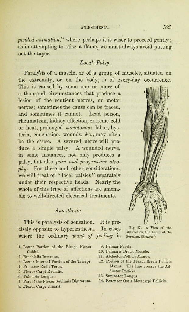
Fig. 87. A View of the
Muscles on the Front of the
where the ordinary want of feeling is Forearm, (Flexors.)
1. Lower Portion of the Biceps Flexor
Cubiti.
2. Brachialis Interims.
3. Lower Internal Portion of the Triceps.
4. Pronator Radii Teres.
5. Flexor Carpi Radialis.
6. Palmaris Longus.
7. Part of the Flexor Sublimis Digitorum.
8. Flexor Carpi XJlnaris.
9. Palmar Fascia.
10. Palmaris Brevis Muscle.
11. Abductor Pollicis Manus.
12. Portion of the Flexor Breyis Pollicis
Manus. The line crosses the Ad-
ductor Pollicis.
13. Supinator Longus.
14. Extensor Ossis Metacarpi Pollicis.