The image contains the following text:
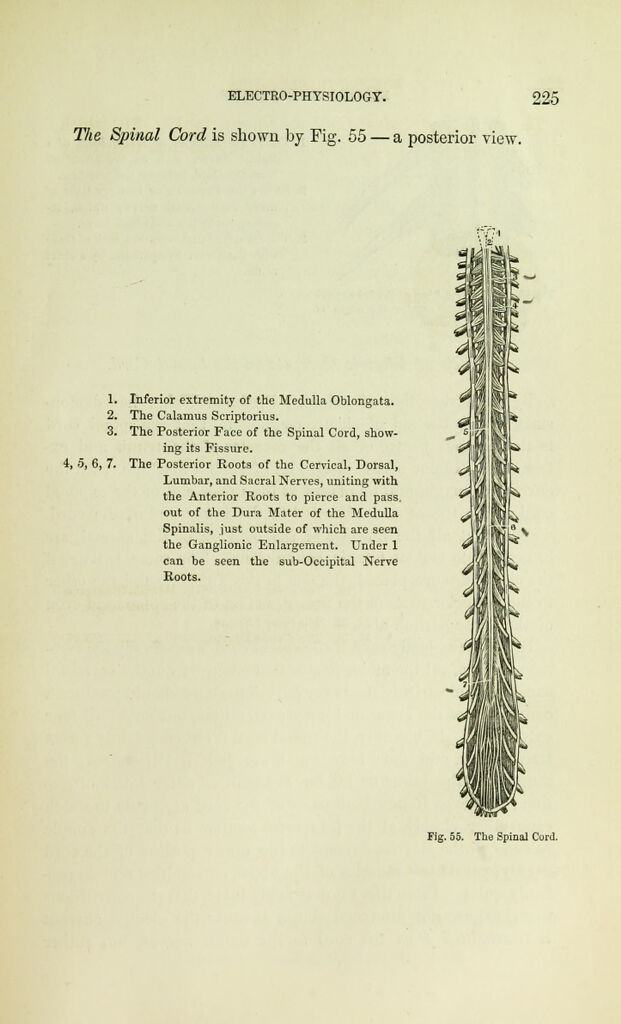
The Spinal Cord is shown by Fig. 55— a posterior view.
1. Inferior extremity of the Medulla Oblongata.
2. The Calamus Scriptorius.
3. The Posterior Face of the Spinal Cord, show-
ing its Fissure.
i, 0, 6, 7. The Posterior Roots of the Cervical, Dorsal,
Lumbar, and Sacral Nerves, uniting with
the Anterior Roots to pierce and pass.
out of the Dura Mater of the Medulla
Spinalis, just outside of which are seen
the Ganglionic Enlargement. Under 1
can be seen the sub-Occipital Nerve
Roots.