

Electro-physiology and electro-therapeutics : showing the best methods for the medical uses of electricity / By Alfred C. Garratt.
463/740
- Publication/creation
- Boston : Ticknor and Fields, 1861.
- Physical description
- 3 unnumbered pages, 716 pages, 2 leaves of plates : illustrations ; 26 cm
- Contributors
-
Garratt, Alfred C. (Alfred Charles), 1813?-1891
Harvey Cushing/John Hay Whitney Medical Library
- Notes
-
3d ed. has title: Medical electricity
Includes index and testimonials
- Type/technique
- Electronic books
- Subjects
-
Electrophysiology
Electrotherapeutics
- Attribution and usage
-
This material has been provided by the Harvey Cushing/John Hay Whitney Medical Library at Yale University, through the Medical Heritage Library. The original may be consulted at the Harvey Cushing/John Hay Whitney Medical Library at Yale University.
This work has been identified as being free of known restrictions under copyright law, including all related and neighbouring rights and is being made available under the Creative Commons, Public Domain Mark.
You can copy, modify, distribute and perform the work, even for commercial purposes, without asking permission.
The image contains the following text:
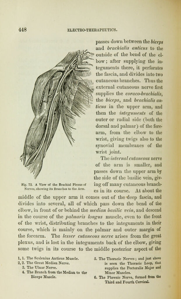
passes down between the biceps
and brachialis anlicus to the
outside of the bend of the el-
bow ; after supplying the in-
teguments there, it perforates
the fascia, and divides into two
cutaneous branches. Thus the
external cutaneous nerve first
supplies the coraco-brachialis,
the biceps, and brachialis an-
ticus in the upper arm, and
then the integuments of the
outer or radial side (both the
dorsal and palmar) of the fore-
arm, from the elbow to the
wrist, giving twigs also to the
synovial membranes of the
wrist joint.
The internal cutaneous nerve
of the arm is smaller, and
passes down the upper arm by
the side of the basilic vein, giv-
ing off many cutaneous brandi-
es in its course. At about the
middle of the upper arm it comes out of the deep fascia, and
divides into several, all of which pass down the bend of the
elbow, in front of or behind the median basilic vein, and descend
in the course of the palmaris longus muscle, even to the front
of the wrist, distributing branches to the integuments in their
course, which is mainly on the palmar and outer margin of
the forearm. The lesser cutaneous nerve arises from the great
plexus, and is lost in the integuments back of the elbow, giving
some twigs in its course to the middle posterior aspect of the
Fig. 72. A View of the Brachial Plexus of
Nerves, showing its Branches to the Arm.
1, 1. The Scalenius Anticus Muscle.
2, 2. The Great Median Nerve.
3. The Ulnar Nerve.
4. The Branch from the Median to the
Biceps Muscle.
5. The Thoracic Nerves ; and just above
is seen the Thoracic Loop, that
supplies the Pectoralis Major and
Minor Muscles.
6. The Phrenic Nerve, formed from the
Third and Fourth Cervical.