The image contains the following text:
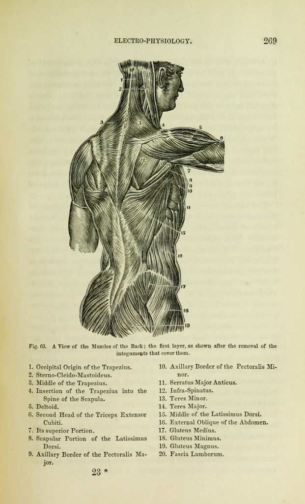
Fig. 63. A View of the Muscles of the Back; the first layer, as shown after the removal of the
integuments that cover them.
1. Occipital Origin of the Trapezius.
2. Sterno-Cleido-Mastoideus.
3. Middle of the Trapezius.
4. Insertion of the Trapezius into the
Spine of the Scapula.
5. Deltoid.
G. Second Head of the Triceps Extensor
Cubiti.
7. Its superior Portion.
8. Scapular Portion of the Latissimus
Dorsi.
9. Axillary Border of the Pectoralis Ma-
jor.
23 *
10. Axillary Border of the Pectoralis Mi-
nor.
11. Serratus Major Anticus.
12. Infra-Spinatus.
13. Teres Minor.
14. Teres Major.
15. Middle of the Latissimus Dorsi.
16. External Oblique of the Abdomen.
17. Gluteus Medius.
18. Gluteus Minimus.
19. Gluteus Magnus.
20. Fascia Lumborum.