

Electro-physiology and electro-therapeutics : showing the best methods for the medical uses of electricity / By Alfred C. Garratt.
235/740
- Publication/creation
- Boston : Ticknor and Fields, 1861.
- Physical description
- 3 unnumbered pages, 716 pages, 2 leaves of plates : illustrations ; 26 cm
- Contributors
-
Garratt, Alfred C. (Alfred Charles), 1813?-1891
Harvey Cushing/John Hay Whitney Medical Library
- Notes
-
3d ed. has title: Medical electricity
Includes index and testimonials
- Type/technique
- Electronic books
- Subjects
-
Electrophysiology
Electrotherapeutics
- Attribution and usage
-
This material has been provided by the Harvey Cushing/John Hay Whitney Medical Library at Yale University, through the Medical Heritage Library. The original may be consulted at the Harvey Cushing/John Hay Whitney Medical Library at Yale University.
This work has been identified as being free of known restrictions under copyright law, including all related and neighbouring rights and is being made available under the Creative Commons, Public Domain Mark.
You can copy, modify, distribute and perform the work, even for commercial purposes, without asking permission.
The image contains the following text:
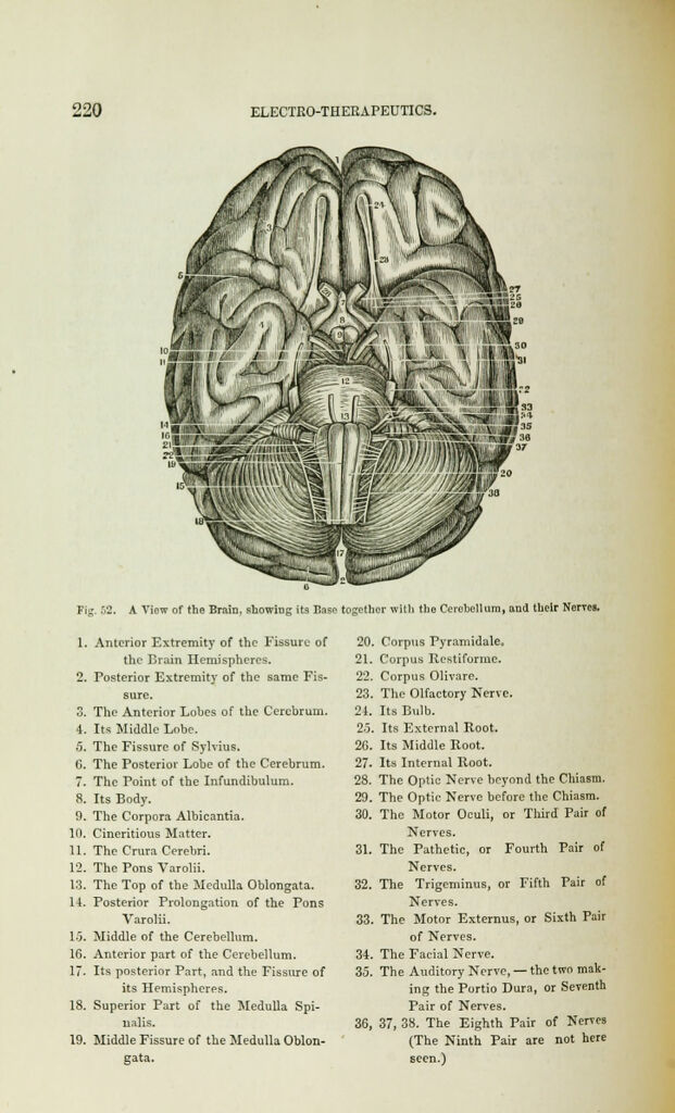
Fig. 52. A View of the Brain, showing its Base together with the Cerebellum, and their Nerves.
1. Anterior Extremity of the Fissure of 20.
the Brain Hemispheres. 21.
2. Posterior Extremity of the same Fis- 22.
sure. 23.
3. The Anterior Lobes of the Cerebrum. 24.
4. Its Middle Lobe. 2.5.
5. The Fissure of Sylvius. 26.
6. The Posterior Lobe of the Cerebrum. 27.
7. The Point of the Infundibulum. 28.
8. Its Body. 29.
9. The Corpora Albicantia. 30.
10. Cineritious Matter.
11. The Crura Cerebri. 31.
12. The Pons Varolii.
13. The Top of the Medulla Oblongata. 32.
14. Posterior Prolongation of the Pons
Varolii. 33.
1.3. Middle of the Cerebellum.
16. Anterior part of the Cerebellum. 34.
17- Its posterior Part, and the Fissure of 35.
its Hemispheres.
18. Superior Part of the Medulla Spi-
nalis. 36,
19. Middle Fissure of the Medulla Oblon-
gata.
Corpus Pyramidale0
Corpus Restiformc.
Corpus Olivare.
The Olfactory Nerve.
Its Bulb.
Its External Root.
Its Middle Root.
Its Internal Root.
The Optic Nerve beyond the Chiasm.
The Optic Nerve before the Chiasm.
The Motor Oculi, or Third Pair of
Nerves.
The Pathetic, or Fourth Pair of
Nerves.
The Trigeminus, or Fifth Pair of
Nerves.
The Motor Externus, or Sixth Pair
of Nerves.
The Facial Nerve.
The Auditory Nerve, — the two mak-
ing the Portio Dura, or Seventh
Pair of Nerves.
37, 38. The Eighth Pair of Nerves
(The Ninth Pair are not here
seen.)