

Electro-physiology and electro-therapeutics : showing the best methods for the medical uses of electricity / By Alfred C. Garratt.
177/740
- Publication/creation
- Boston : Ticknor and Fields, 1861.
- Physical description
- 3 unnumbered pages, 716 pages, 2 leaves of plates : illustrations ; 26 cm
- Contributors
-
Garratt, Alfred C. (Alfred Charles), 1813?-1891
Harvey Cushing/John Hay Whitney Medical Library
- Notes
-
3d ed. has title: Medical electricity
Includes index and testimonials
- Type/technique
- Electronic books
- Subjects
-
Electrophysiology
Electrotherapeutics
- Attribution and usage
-
This material has been provided by the Harvey Cushing/John Hay Whitney Medical Library at Yale University, through the Medical Heritage Library. The original may be consulted at the Harvey Cushing/John Hay Whitney Medical Library at Yale University.
This work has been identified as being free of known restrictions under copyright law, including all related and neighbouring rights and is being made available under the Creative Commons, Public Domain Mark.
You can copy, modify, distribute and perform the work, even for commercial purposes, without asking permission.
The image contains the following text:
be found complete in the structure of the brain ; for it is now
manifest that almost every structure and arrangement here
found may be nearly imitated by voltaic arrangements, combi-
nations, and manifestations.
Muscular motion consists in a change in the arrangement and
composition of tbe matter contained within the ultimate fibres,
so that they become shorter, and consequently thicker and wider
in diameter ; and simply tins is contraction, and this latter moves
the limbs. Nerves are distributed to all the muscles ; but this
supply is in a very unequal degree. The termination of the larger
muscle nerves appears often to abut on many muscle fibres, and
also to be in loops, running likewise transversely to the general
course of the muscle fibre. Each of
these muscle fibres is completely en-
veloped by the minute blood vessels
which run parallel with them. This
admirable arrangement of the capil-
laries yields the abundant supply of
bright arterial blood, so necessary for
the manifestation of muscular motion ;
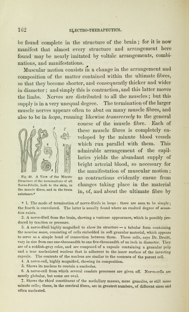
Fig. 48. a view of the Minute as contractions evidently ensue from
Structure of the terminations of all
Nerve-Fibrils, both to the skin, in changes taking place in the material
the moscle fibres, and in the brain • f d a)jout tj ultimate fibre by
Bulitttance.* J
* 1. The mode of termination of nerve-fibrils in loops : three are seen to be simple;
the fourth is convoluted. The latter is usually found where an exalted degree of sensa-
tion exists.
2. A nerve-fibril from the brain, showing a varicose appearance, which is possibly pro-
duced by traction or pressure.
3. A nerve-fibril highly magnified to show its structure — a tubular form containing
the ncurine mass, consisting of cells embedded in soft granular material, which appears
to serve as a simple bond of connection between them. These cells, says Dr. Druitt,
vary in size from one one-thousandth to one five-thousandth of an inch in diameter. They
are of a reddish-gray color, and are composed of a capsule containing a granular pulp
and a true nucleolated nucleus that is adherent to the inner surface of the investing
capsule. The contents of the nucleus are similar to the contents of the parent cell.
4. A nerve-cell, highly magnified, showing its composition.
5. Shows its nucleus to contain a nucleolus.
6. A nerve-cell from which several caudate processes are given off. Nerve-cells are
mostly globular, but some are oval.
7. Shows the third constituent of the medullary masses, mere granules, or still more
minute cells; these, in the cerebral fibres, are in greatest numbers, of different sizes and
often nucleated.