

Electro-physiology and electro-therapeutics : showing the best methods for the medical uses of electricity / By Alfred C. Garratt.
268/740
- Publication/creation
- Boston : Ticknor and Fields, 1861.
- Physical description
- 3 unnumbered pages, 716 pages, 2 leaves of plates : illustrations ; 26 cm
- Contributors
-
Garratt, Alfred C. (Alfred Charles), 1813?-1891
Harvey Cushing/John Hay Whitney Medical Library
- Notes
-
3d ed. has title: Medical electricity
Includes index and testimonials
- Type/technique
- Electronic books
- Subjects
-
Electrophysiology
Electrotherapeutics
- Attribution and usage
-
This material has been provided by the Harvey Cushing/John Hay Whitney Medical Library at Yale University, through the Medical Heritage Library. The original may be consulted at the Harvey Cushing/John Hay Whitney Medical Library at Yale University.
This work has been identified as being free of known restrictions under copyright law, including all related and neighbouring rights and is being made available under the Creative Commons, Public Domain Mark.
You can copy, modify, distribute and perform the work, even for commercial purposes, without asking permission.
The image contains the following text:
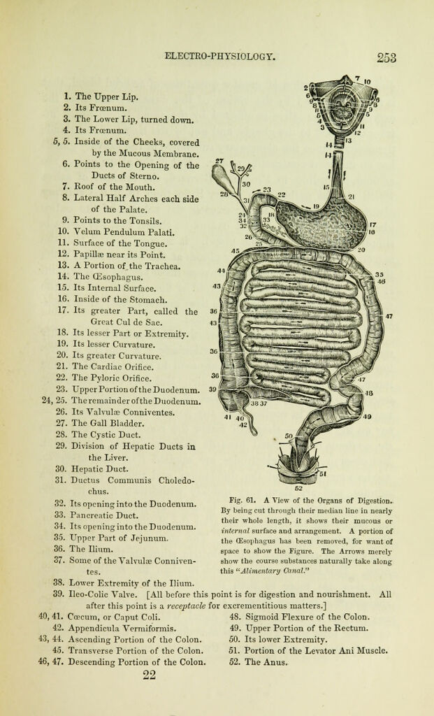
1. The Upper Lip.
2. Its Frcenum.
3. The Lower Lip, turned down.
4. Its Froenum.
5, 5. Inside of the Cheeks, covered
by the Mucous Membrane.
6. Points to the Opening of the
Ducts of Sterno.
7. Roof of the Mouth.
8. Lateral Half Arches each side
of the Palate.
9. Points to the Tonsils.
10. Velum Pendulum Palati.
11. Surface of the Tongue.
12. Papilla; near its Point.
13. A Portion of the Trachea.
14. The Oesophagus.
15. Its Internal Surface.
16. Inside of the Stomach.
17. Its greater Part, called the
Great Cul de Sac.
18. Its lesser Part or Extremity.
19. Its lesser Curvature.
20. Its greater Curvature.
21. The Cardiac Orifice.
22. The Pyloric Orifice.
23. Upper Portion of the Duodenum.
24, 2.5. TheremainderoftheDuodenum.
26. Its Valvulce Conniventes.
27. The Gall Bladder.
28. The Cystic Duct.
29. Division of Hepatic Ducts in
the Liver.
30. Hepatic Duct.
31. Ductus Communis Choledo-
chus.
32. Its opening into the Duodenum.
33. Pancreatic Duct.
34. Its opening into the Duodenum.
35. Upper Part of Jejunum.
36. The Ilium.
37. Some of the Valvulse Conniven-
tes.
38. Lower Extremity of the Ilium.
39. Ileo-Colic Valve. [All before this
after this point is a receptacle
40, 41. Ccrcum, or Caput Coli.
42. Appendicula Vermiformis.
43, 44. Ascending Portion of the Colon.
45. Transverse Portion of the Colon.
46, 47. Descending Portion of the Colon.
22
Fig. 61. A View of the Organs of Digestion.
By being cut through their median line in nearly
their whole length, it shows their mucous or
internal surface and arrangement. A portion of
the (Esophagus has been removed, for want of
space to show the Figure. The Arrows merely
show the course substances naturally take along
this "Alimentary Canal."
point is for digestion and nourishment. All
for excrementitious matters.]
48. Sigmoid Flexure of the Colon.
49. Upper Portion of the Rectum.
50. Its lower Extremity.
51. Portion of the Levator Ani Muscle.
52. The Anus.