The image contains the following text:
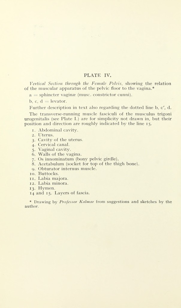
Vertical Section through the Female Pelvis, showing the relation
of the muscular apparatus of the pelvic floor to the vagina.*
a = sphincter vaginae (muse, constrictor cunni).
b, c, d = levator.
Further description in text also regarding the dotted line b, c', d.
The transverse-running muscle fasciculi of the museums trigoni
urogenitalis (see Plate I.) are for simplicity not drawn in, but their
position and direction are roughly indicated by the line 15.
1. Abdominal cavity.
2. Uterus.
3. Cavity of the uterus.
4. Cervical canal.
5. Vaginal cavity.
6. Walls of the vagina.
7. Os innominatum (bony pelvic girdle).
8. Acetabulum (socket for top of the thigh bone).
9. Obturator internus muscle.
10. Buttocks.
11. Labia majora.
12. Labia minora.
13. Hymen.
14 and 15. Layers of fascia.
* Drawing by Professor Kalmar from suggestions and sketches by the
author.