Sex efficiency through exercises : special physical culture for women / by Th. H. van de Velde ; [photos, by E. Steinemann].
233/426
- (canvas 234)
The image contains the following text:
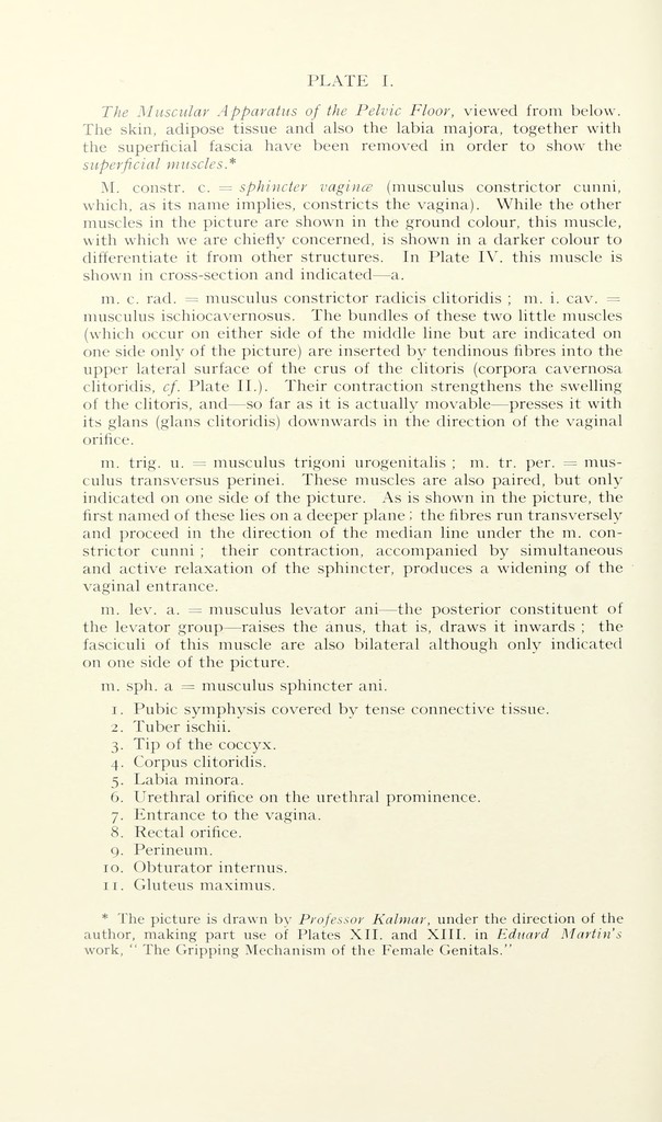
The Muscular Apparatus of the Pelvic Floor, viewed from below.
The skin, adipose tissue and also the labia majora, together with
the superficial fascia have been removed in order to show the
superficial muscles*
M. constr. c. = sphincter vagince (musculus constrictor cunni,
which, as its name implies, constricts the vagina). While the other
muscles in the picture are shown in the ground colour, this muscle,
with which we are chiefly concerned, is shown in a darker colour to
differentiate it from other structures. In Plate IV. this muscle is
shown in cross-section and indicated—a.
m. c. rad. = musculus constrictor radicis clitoridis ; m. i. cav. =
musculus ischiocavernosus. The bundles of these two little muscles
(which occur on either side of the middle line but are indicated on
one side only of the picture) are inserted by tendinous fibres into the
upper lateral surface of the crus of the clitoris (corpora cavernosa
clitoridis, cf. Plate II.). Their contraction strengthens the swelling
of the clitoris, and—so far as it is actually movable—presses it with
its glans (glans clitoridis) downwards in the direction of the vaginal
orifice.
m. trig. u. = musculus trigoni urogenitalis ; m. tr. per. = mus-
culus transversus perinei. These muscles are also paired, but only
indicated on one side of the picture. As is shown in the picture, the
first named of these lies on a deeper plane ; the fibres run transversely
and proceed in the direction of the median line under the m. con-
strictor cunni ; their contraction, accompanied by simultaneous
and active relaxation of the sphincter, produces a widening of the
vaginal entrance.
m. lev. a. = musculus levator ani—the posterior constituent of
the levator group—raises the anus, that is, draws it inwards ; the
fasciculi of this muscle are also bilateral although only indicated
on one side of the picture.
m. sph. a = musculus sphincter ani.
1. Pubic symphysis covered by tense connective tissue.
2. Tuber ischii.
3. Tip of the coccyx.
4. Corpus clitoridis.
5. Labia minora.
6. Urethral orifice on the urethral prominence.
7. Entrance to the vagina.
8. Rectal orifice.
9. Perineum.
10. Obturator internus.
11. Gluteus maximus.
* The picture is drawn by Professor Kalmar, under the direction of the
author, making part use of Plates XII. and XIII. in Editard Martin's
work, " The Gripping Mechanism of the Female Genitals."