Electro-physiology and electro-therapeutics : showing the best methods for the medical uses of electricity / By Alfred C. Garratt.
483/740
468 (canvas 484)
The image contains the following text:
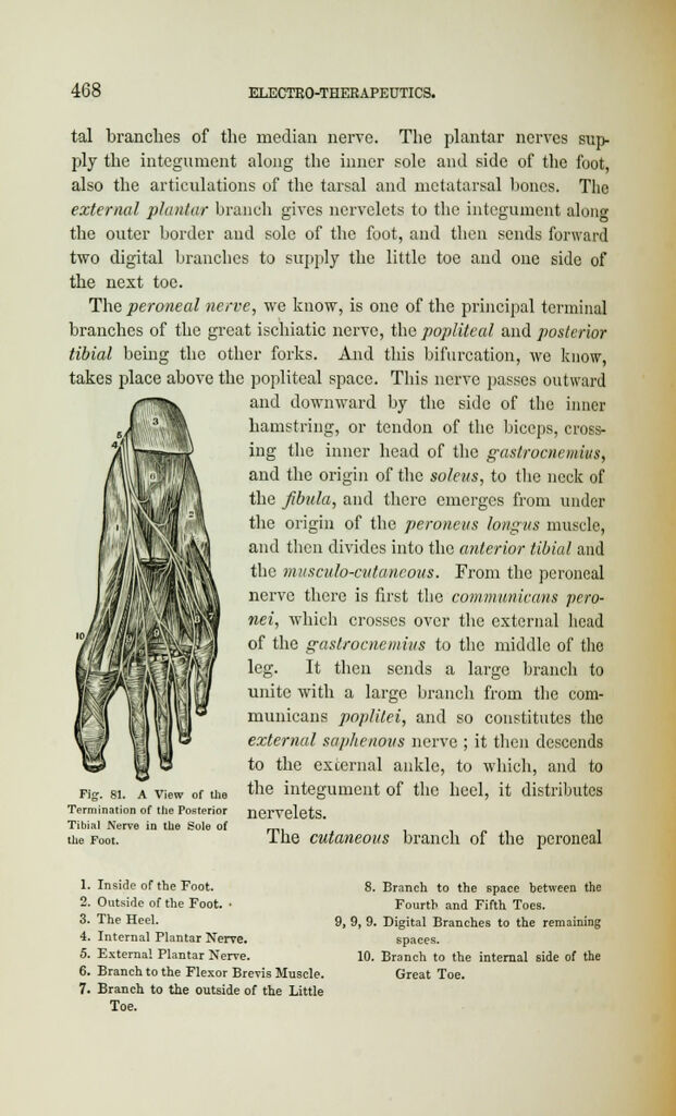
tal branches of the median nerve. The plantar nerves sup-
ply the integument along the inner sole and side of the foot,
also the articulations of the tarsal and metatarsal bones. The
external plantar branch gives nervelets to the integument along
the outer border and sole of the foot, and then sends forward
two digital branches to supply the little toe and one side of
the next toe.
The peroneal nerve, we know, is one of the principal terminal
branches of the great ischiatic nerve, the popliteal and posterior
tibial being the other forks. And this bifurcation, we know,
takes place above the popliteal space. This nerve passes outward
and downward by the side of the inner
hamstring, or tendon of the biceps, cross-
ing the inner head of the gastrocnemius,
and the origin of the solens, to the neck of
the fibula, and there emerges from under
the origin of the peroneus longus muscle,
and then divides into the anterior tibial and
the mitsculo-cutancous. From the peroneal
nerve there is first the communicans pero-
nei, which crosses over the external head
of the gastrocnemius to the middle of the
leg. It then sends a large branch to
unite with a large branch from the com-
municans poplitei, and so constitutes the
external saphenous nerve ; it then descends
to the external ankle, to which, and to
Fig. si. a view or ti the integument of the heel, it distributes
Termination of the Posterior nervelets.
Tibial Nerve in the Sole of mi
ihe Foot. i he cutaneous branch of the peroneal
1. Inside of the Foot.
2. Outside of the Foot. •
3. The Heel.
4. Internal Plantar Nerve.
5. Externa! Plantar Nerve.
6. Branch to the Flexor Brevis Muscle.
7. Branch to the outside of the Little
Toe.
8. Branch to the space between the
Fourth and Fifth Toes.
9, 9, 9. Digital Branches to the remaining
spaces.
10. Branch to the internal side of the
Great Toe.