Electro-physiology and electro-therapeutics : showing the best methods for the medical uses of electricity / By Alfred C. Garratt.
248/740
233 (canvas 249)
The image contains the following text:
current is opened, the contraction of the iris as instantly ceases.
If the small sponge tipped ivory electrodes are now applied to
the opposite edges of the cornea of one eye, or a little farther
hack on the sclerotica of the eye, then the radiar fibres of the
iris are extended, as is an extensor muscle, and thus the pupil
is enlarged. If these little electrodes are applied to the right
and left of the eyeball, the pupil then assumes the form of an
egg standing upon end; if the electrodes touch the eyeball
above and below, then the oval pupil is horizontal. Thus we
can act on the iris of the eye by the electric current so as to
contract or dilate the pupil according as we direct the electrodes
to the sphincter pupillw, or to the dilator pupillm. The pupil of
the eye in man can thus be constricted simply by means of a
single voltaic pair, as by my local silver battery, if the one metal
is placed in the nostril, while the other is upon the tongue; and
then — the patient sitting in the mildest light of the room, where
there is only just light enough to see the oscillations of the
pupil, — upon making and breaking contact between the two
metals by the clasp of the insulated conductor, the pupil is seen
to dilate and contract; or, rather, to contract with the contact,
and to dilate with the breaking of the circuit.
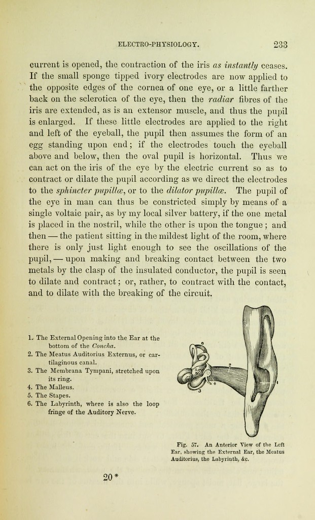
l.i The External Opening into the Ear at the
bottom of the Concha.
2. The Meatus Auditorius Externus, or car-
tilaginous canal.
3. The Membrana Tympani, stretched upon
its ring.
4. The Malleus.
5. The Stapes.
6. The Labyrinth, where is also the loop
fringe of the Auditory Nerve.
Fig. 57. An Anterior View of the Left
Ear, showing the External Ear, the Meatus
Auditorius, the Labyrinth, &c.
20'