Electro-physiology and electro-therapeutics : showing the best methods for the medical uses of electricity / By Alfred C. Garratt.
262/740
247 (canvas 263)
The image contains the following text:
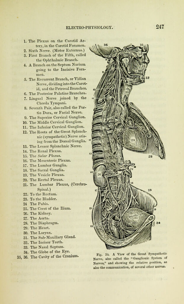
1. The Plexus on the Carotid Ar-
tery, in the Carotid Foramen.
2. Sixth Nerve, (Motor Externus.)
3. First Branch of the Fifth, called
the Ophthalmic Branch.
4. A Branch on the Septum Narium
going to the Incisive Fora-
men.
5. The Recurrent Branch, or Vidian
Nerve, dividing into the Carot-
id, and the Petrosal Branches.
6. The Posterior Palatine Branches.
7. Lingual Nerve joined by the
Chorda Tympani.
8. Seventh Pair, also called the Por-
tio Dura, or Facial Nerve.
9. The Superior Cervical Ganglion.
10. The Middle Cervical Ganglion.
11. The Inferior Cervical Ganglion.
12. The Roots of the Great Splanch-
nic (sympathetic) Nerve aris-
ing from the Dorsal Ganglia.
13. The Lesser Splanchnic Nerve.
14. The Renal Plexus.
15. The Solar Plexus.
16. The Mesenteric Plexus.
17. The Lumbar Ganglia.
18. The Sacral Ganglia.
19. The Vesicle Plexus.
20. The Rectal Plexus.
21. The Lumbar Plexus, (Cerebro-
spinal.)
22. To the Rectum.
23. To the Bladder.
24 The Pubis.
25. The Crest of the Ilium.
26. The Kidney.
27. The Aorta.
28. The Diaphragm.
29. The Heart.
30. The Larynx.
31. The Sub-Maxillary Gland.
32. The Incisor Teeth.
33. The Nasal Septum.
34. The Globe of the Eye.
35, 36. The Cavity of the Cranium.
Fig. 59. A View of the Great Sympathetic
Nerve, also called the "Ganglionic System of
Nerves," and Bhowing the relative position, as
also the communication, of Beveral other nerves.